


 HOME
HOME


검사실 효율성 향상
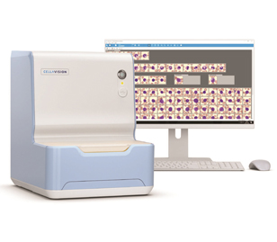
최소한의 공간을 차지하는 컴팩트한 사이즈 (W280xD390xH370mm)로 검사실 공간을 효율적으로 사용할 수 있으며, 네트워크 시스템이 구축된 검사실과 실시간으로 이미지 분석 결과를 확인할 수 있습니다.
Digital Scan 기능
DC-1은 도말된 슬라이드에서 세포의 디지털 이미지를 캡쳐한 후 WBC, RBC, PLT를 자동 분석하여 결과를 제공하며, 디지털 스캔 기능도 제공하므로 다양한 슬라이드에 여러 목적으로 활용할 수 있습니다.
분석항목
1) 백혈구 분류(WBC Classification)
Segmented and band neutrophils, eosinophils, basophils, lymphocytes, monocytes, blast cells, promyeolocytes, myelocytes, metamyelocytes, variant lymphocytes, plasma cells.
2) 비 백혈구 분류(non-WBC Classification)
Smudge artifacts, giant platelets, platelet clumps, erythroblasts(NRBC), unidentified
3) 적혈구 전단계 분류(RBC pre-characterization)
Automated pre-characterization of aniso-, micro- and macrocytosis, polychromasia, hypochromasia, poikilocytosis
*8 high power fields (100배율)에서 수행
4) 혈소판 예측 (PLT Estimate)
8 high power fields (100배율)에서 혈소판 농도의 매뉴얼 예측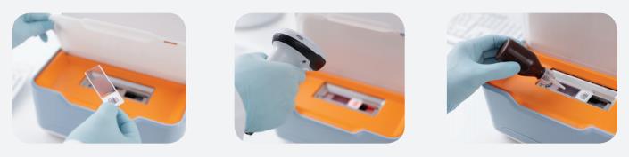 Step 1. 염색된 도말 표본을 분석기의 로드 트레이에 놓습니다.
Step 2. 수동 또는 Option 사양의 바코드 리더기로 슬라이드의 Order ID를 입력합니다.
Step 3. 슬라이드에 Immersion oil 점적하고 해치를 닫은 다음, Processing을 시작합니다.
Segmented and band neutrophils, eosinophils, basophils, lymphocytes, monocytes, blast cells, promyeolocytes, myelocytes, metamyelocytes, variant lymphocytes, plasma cells.
2) 비 백혈구 분류(non-WBC Classification)
Smudge artifacts, giant platelets, platelet clumps, erythroblasts(NRBC), unidentified
3) 적혈구 전단계 분류(RBC pre-characterization)
Automated pre-characterization of aniso-, micro- and macrocytosis, polychromasia, hypochromasia, poikilocytosis
*8 high power fields (100배율)에서 수행
4) 혈소판 예측 (PLT Estimate)
8 high power fields (100배율)에서 혈소판 농도의 매뉴얼 예측
Step 2. 수동 또는 Option 사양의 바코드 리더기로 슬라이드의 Order ID를 입력합니다.
Step 3. 슬라이드에 Immersion oil 점적하고 해치를 닫은 다음, Processing을 시작합니다.
